Home
/ Shoulder Joint Anatomy Diagram Easy - Shoulder Joint Type Articular Surfaces Capsule And Ligaments Relations Movements And Applie Shoulder Joint Shoulder Joint Anatomy Acromioclavicular Joint - Lumbar spine anatomy is unique in being strong and flexible.
Shoulder Joint Anatomy Diagram Easy - Shoulder Joint Type Articular Surfaces Capsule And Ligaments Relations Movements And Applie Shoulder Joint Shoulder Joint Anatomy Acromioclavicular Joint - Lumbar spine anatomy is unique in being strong and flexible.
Shoulder Joint Anatomy Diagram Easy - Shoulder Joint Type Articular Surfaces Capsule And Ligaments Relations Movements And Applie Shoulder Joint Shoulder Joint Anatomy Acromioclavicular Joint - Lumbar spine anatomy is unique in being strong and flexible.. The shoulder joint by quan fu gan 67157 views. Click now and learn everything about its anatomy and function at kenhub! 1 this mobility provides the upper extremity with tremendous range of motion such as adduction, abduction, flexion, extension, internal rotation, external rotation, and 360° circumduction in. Use the mouse scroll wheel to move the images up and down alternatively use the tiny arrows (>>) on both side of the image to move the images. The shoulder joint is the connection between the chest and the upper extremity.
8 name the arteries and the nerves that supply shoulder joint. Equally extensive are the muscles affecting the shoulder movement, including: The shoulder joint (glenohumeral joint) is a ball and socket joint between the scapula and the the shoulder joint is formed by the articulation of the head of the humerus with the glenoid cavity (or in this article, we shall look at the anatomy of the shoulder joint and its important clinical correlations. Chronic or acute wear and tear on the. Set of human joints, elbow, knee joint, hip and shoulder joint, skeletal bone structure.
Easy Notes On Shoulder Glenohumeral Joint Learn In Just 3 Mins Earth S Lab from www.earthslab.com Robin smithuis and henk jan van der woude. In human anatomy, the shoulder joint comprises the part of the body where the humerus attaches to the scapula.1 there are two kinds of cartilage in the joint. Set of human joints, elbow, knee joint, hip and shoulder joint, skeletal bone structure. 8 name the arteries and the nerves that supply shoulder joint. All about the shoulder muscles. Click now and learn everything about its anatomy and function at kenhub! Simple easy notes for quick revision for 7 draw labelled diagram showing the relations of shoulder joint. Moderate to serve pain along the outer edge of the clavicle, when raising arm over the head, and/or when reaching arm across the body.
This incongruent bony anatomy allows for the wide range of movement available at the shoulder joint but is also the reason for the lack of joint stability.
Skeleton of an arm, vintage engraved illustration. Home > blog > anatomy > shoulder anatomy: Click now and learn everything about its anatomy and function at kenhub! Normal anatomy, variants and checklist. The shoulder joint is vulnerable to dislocations from sudden jerks of the arm, especially in children before strong muscles have developed. Due to the tension by the anterior band of the inferior ghl labral teras will be easier to detect. The shoulder joint is the most mobile joint in the human body and responsible for movements of arm and scapula. Chronic or acute wear and tear on the. The shoulder joint is formed where the humerus (upper arm bone) fits into the scapula. Describe the structure of the shoulder should begin with bone parts that include: Labeled human shoulder bone anatomical vector illustration diagram poster. Learn vocabulary, terms and more with flashcards, games and other study tools. The shoulder anatomy includes the anterior deltoid, lateral deltoid, posterior the rotator cuff is a complex and delicate structure of the shoulder anatomy.
Posted on december 13, 2018december 12, 2018. Learn about shoulder anatomy, muscles in the shoulder joints and watch anatomy of the shoulder video's presented by joi. Equally extensive are the muscles affecting the shoulder movement, including: The shoulder joint is the connection between the chest and the upper extremity. How to draw heart diagram in exams ?
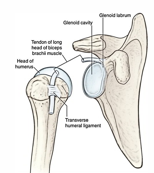
Shoulder Joint Type Articular Surfaces Capsule And Ligaments Relations Movements And Applie Shoulder Joint Shoulder Joint Anatomy Acromioclavicular Joint from i.pinimg.com The glenohumearal joint has a greater range of motion than any other joint in the body. 8 name the arteries and the nerves that supply shoulder joint. • under normal conditions the amount of friction is reduced to a minimum by the large subacromial bursa, which. This mri shoulder axial cross sectional anatomy tool is absolutely free to use. Dislocation of the shoulder is extremely painful and may require surgical repair or even cause permanent damage. The human shoulder is the most mobile joint in the body. As the disease progresses, night pain becomes more common. Three bones come together at the shoulder joint.
Use the mouse scroll wheel to move the images up and down alternatively use the tiny arrows (>>) on both side of the image to move the images.
Normal anatomy, variants and checklist. The first type is the white cartilage on the ends of the bones (called articular cartilage) which allows the bones to glide and move on each other. Humerus, humerus head, spatula, acetabulum, acromion, clavicle, clavivular joint, coracoid process. Shoulder anatomy is an elegant piece of machinery having the greatest range of motion of any joint in the deepest layer of the shoulder includes the bones and the joints. Describe the structure of the shoulder should begin with bone parts that include: Webmd's shoulder anatomy page provides an image of the parts of the shoulder and describes its function, shoulder problems, and more. Just remember the articulating surfaces. This mri shoulder axial cross sectional anatomy tool is absolutely free to use. Various types of injuries and degenerative conditions can cause the shoulder to become painful. • during abduction of the shoulder joint, the supraspinatus tendon is exposed to friction against the acromion. The students must thoroughly study the shoulder joint as it usually undergoes recurrent dislocations and is the most common joint to dislocate. You can see it enclosing the glenohumeral joint and you can see its attachment on the anatomical neck that's the shoulder joint. Home > blog > anatomy > shoulder anatomy:
Erythrocyte sedimentation rate (esr) by shabab ali 21093 views. Equally extensive are the muscles affecting the shoulder movement, including: The shoulder joint is formed where the humerus (upper arm bone) fits into the scapula. Chronic or acute wear and tear on the. You can see it enclosing the glenohumeral joint and you can see its attachment on the anatomical neck that's the shoulder joint.
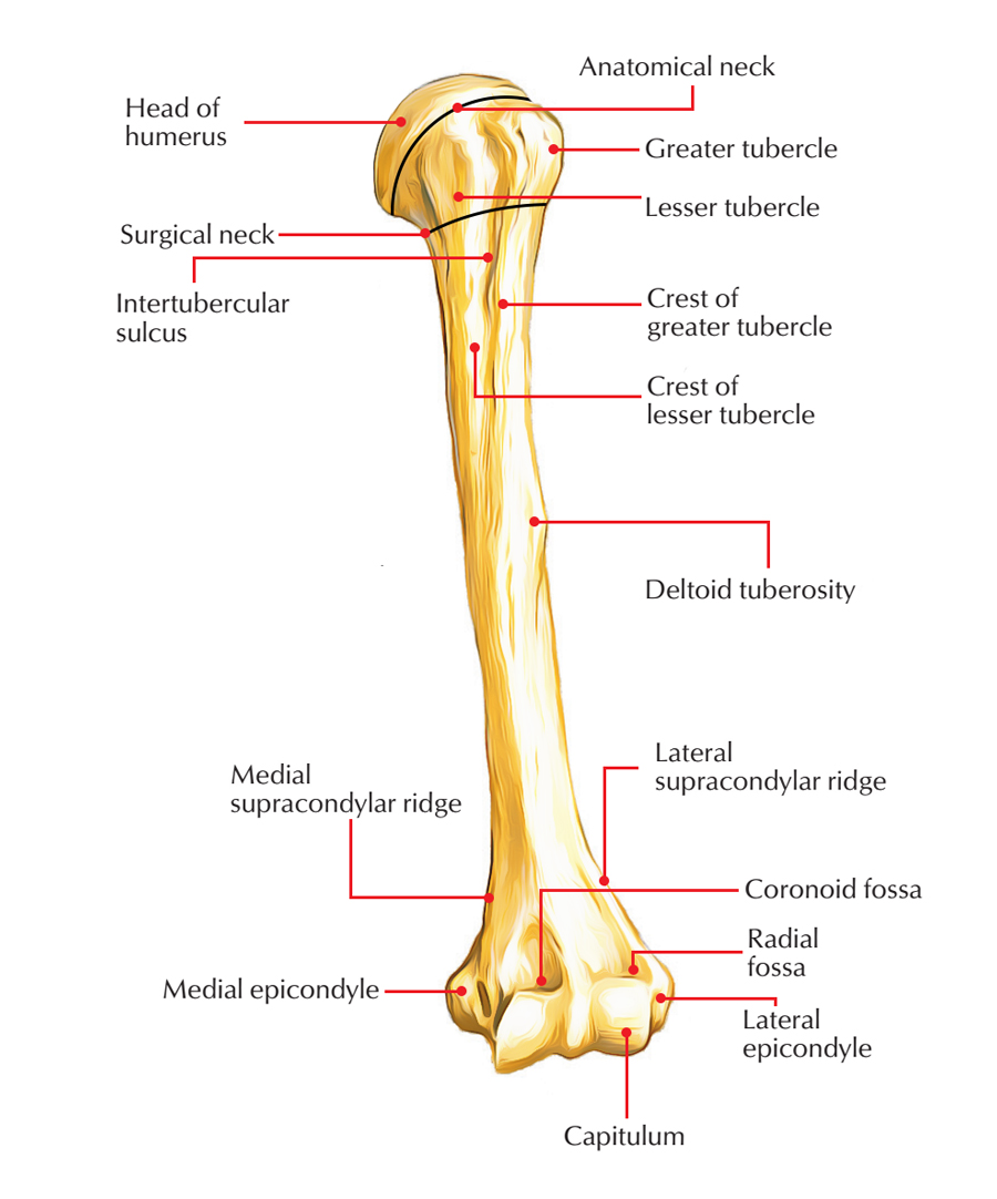
Humeral Anatomy Anatomy Drawing Diagram from www.earthslab.com Three bones come together at the shoulder joint. This incongruent bony anatomy allows for the wide range of movement available at the shoulder joint but is also the reason for the lack of joint stability. Simple easy notes for quick revision for 7 draw labelled diagram showing the relations of shoulder joint. The next layer is made up of the when you realize all the different ways and positions we use our hands every day, it is easy to. Various types of injuries and degenerative conditions can cause the shoulder to become painful. Just remember the articulating surfaces. Shoulder joint is the most mobile joint of the human body. • during abduction of the shoulder joint, the supraspinatus tendon is exposed to friction against the acromion.
Labeled human shoulder bone anatomical vector illustration diagram poster.
Lumbar spine anatomy is unique in being strong and flexible. Shoulder surgery recovery shoulder anatomy joint replacement shoulder injuries knee surgery rotator cuff. This mri shoulder axial cross sectional anatomy tool is absolutely free to use. The glenohumeral joint (shoulder joint) is a synovial ball and socket articulation anatomy ▶ upper limb ▶ joints ▶ shoulder joint (glenohumeral joint). Chronic or acute wear and tear on the. The first type is the white cartilage on the ends of the bones (called articular cartilage) which allows the bones to glide and move on each other. The shoulder joint is formed where the humerus (upper arm bone) fits into the scapula. The students must thoroughly study the shoulder joint as it usually undergoes recurrent dislocations and is the most common joint to dislocate. Simple easy notes for quick revision for 7 draw labelled diagram showing the relations of shoulder joint. • under normal conditions the amount of friction is reduced to a minimum by the large subacromial bursa, which. Equally extensive are the muscles affecting the shoulder movement, including: Shoulder anatomy is an elegant piece of machinery having the greatest range of motion of any joint in the deepest layer of the shoulder includes the bones and the joints. Posted on december 13, 2018december 12, 2018.
Start studying shoulder joint anatomy shoulder anatomy diagram. Three bones come together at the shoulder joint.
